This study used the GrandARM to make a tomogram of a 3D quantum dot superlattice film. It’s state-of-the-art tomography using a full-tilt needle sample, with 0.65 nm resolution over a very large volume of 4.3 x 10⁵ nm3.
The full article can be found at J. Mater. Chem. A, 2020,8, 21339-21339 titled “Structural characterization of a polycrystalline epitaxially-fused colloidal quantum dot superlattice by electron tomography”
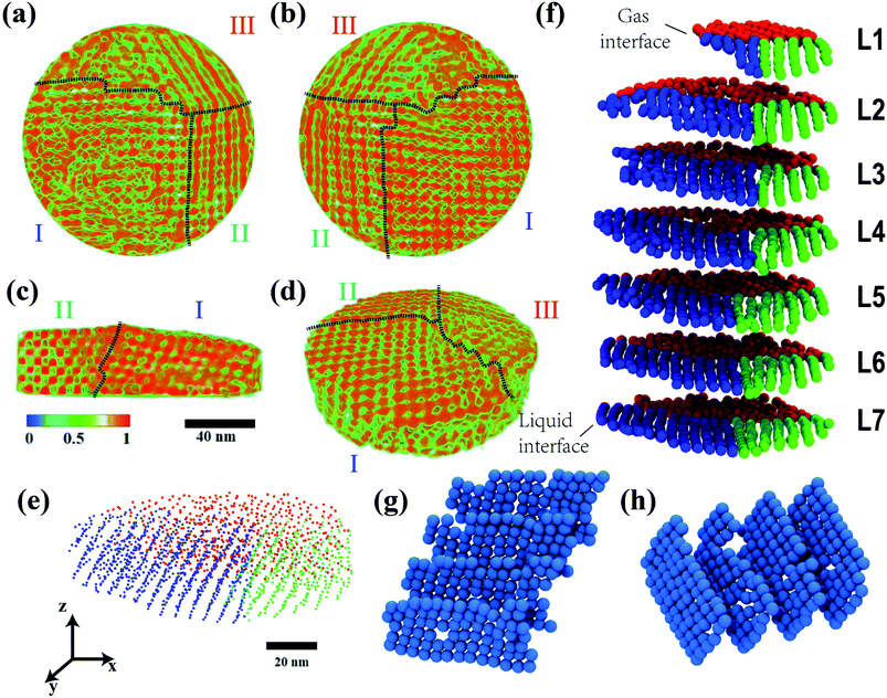

Abstract: Three dimensional epitaxially-fused colloidal quantum dot (QD) superlattices (epi-SLs) feature exceptional electronic coupling and spatial order and are promising systems for studying the emergence of delocalized states and mini-band charge transport in self-assembled solids. However, energy disorder arising from structural defects and aperiodicity has so far resulted in charge carrier localization and slow hopping transport. Detailed 3D structural characterization is critical for rationally improving epi-SL structural perfection to trigger the formation of mini-bands. Here, we analyze the 3D structure of a 120 38 nm disc-shaped region of a PbSe QD epi-SL using full-tilt high-angle annular dark-field electron tomography. The high spatial resolution of the tomographic reconstruction (0.65 nm) enables determination of the center-of-mass coordinates of all 1846 QDs in the sample as well as the size and shape of the thousands of epitaxial connections (necks) between the QDs. The tomogram reveals the detailed crystallography and internal positional disorder of the three SL grains that constitute this sample. A map of the neck network is used to quantify relationships between neck number (the number of necks each QD possesses), average neck diameter, QD location in the film, and the nearest neighbor inter-QD distance and distance distribution. We find a strong positive correlation between neck number and local spatial order, suggesting that future improvements in neck connectivity are likely to simultaneously enhance the overall structural perfection of the epi-SLs. A kinetic Monte Carlo model is employed to estimate the electron mobility of the tomography sample and assess the impact of grain boundaries on charge transport. Our electron tomography study establishes a baseline for the quantitative statistical analysis of structural defects in 3D QD epi-SLs.